FIELD VETERINARY REPORT FOR MERU FOR THE MONTH OF OCTOBER 2016 Report by Dr Bernard Rono Summary This report describes activities of the Meru veterinary unit in northern Kenya in October 2016
FIELD VETERINARY REPORT FOR MERU FOR THE MONTH OF OCTOBER 2016
Report by Dr Bernard Rono
Summary
This report describes activities of the Meru veterinary unit in northern Kenya in October 2016. Many areas continue to experience drought due to scarce water and feed resourcesand increased pressure on wildlife health.
During this month the following clinical interventions were carried out: treatment of a white rhino for cutaneous filariasis, a speared elephant was immobilized to remove the spear and treat the resulting wound and a Grevys zebra was immobilized to attend to injury on its leg. An elephant calf with a deformed leg and traumatic wounds was rescued for further treatment at the Nairobi David Sheldrick Wildlife Trust orphanage.
We would like to acknowledge logistical and financial support provided by the David Sheldrick Wildlife Trust to enable timely veterinary intervention on affected wildlife in northern Kenya.
Case #1 Cutaneous filariasis in a White Rhino
Date: 10th October 2016
Species: White rhino
Sex: Female
Age: Adult
Location: Meru national park
History
The warden in charge of rhino in Meru National Park requested for treatment of a white rhino with a skin infection suspected to have been caused by filarial parasites. The wound had grown progressively over one month and treatment was required to prevent wound infection by bacteria.
Cutaneous filariasis, a parasitic skin condition, has been reported in many white rhinos in Meru National Park. Interventions in white rhino include parenteral administration of Ivermectinand antibiotics although the condition is self limiting in the black rhino.
Immobilization, examination and treatment
An initial attempt at foot darting was unsuccessful after the dart failed to discharge its content. Darting was later achieved from a helicopter. For immobilization we used a combination of Captivon® 5mg and azaperone 60mg in 1.5cc DanInject dart syringe with a 2.2 × 60mm needle. Darting site was the left rump and induction time was 7 minutes.


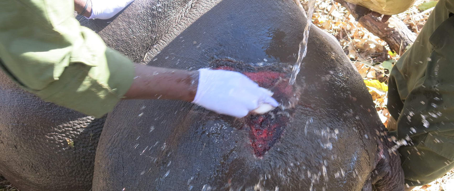
Examination showed ulcerative cutaneous wounds with serrated edges undermined by pockets of pus. Wounds emitted a foul smell due to tissue necrosis caused by bacterial infection. These wounds are characteristic of cutaneous filariasis. The wound was thorough wash with water and dilute hydrogen peroxide before debridement of necrotic tissue.


Tincture of Iodine soaked in gauze swabs was the applied and 1% Ivermectin 300mg administered subcutaneously as well as 20% Oxytetracycline administered intramuscularly.

Reversal
Anesthesia effects were reversed within 3 minutes of intravenous injection of Naltrexone Hcl 150mg.


Prognosis
Filarial wounds respond to ivermectin and antibiotic treatment, therefore this rhino is expected to make a speedy recovery. The rhino monitoring team will observe and report on their progress.
Case #2 Treatment and Rescue of an Elephant Calf
Date: 17th October 2016
Species: Elephant
Sex: Male
Age: Juvenile (less than 3 months)
Location: Meru National Park
History
Tour guides from Elsa’s Kopje reported an elephant calf showing severe lameness on its left hind limb. It was in a herd of 17 elephants and was struggling to keep up with the rest of the herd. Observation showed a deformed limb with a wound on the foot pad. Treatment was required to prevent wound infection.
Immobilsiation, examination and rescue
To capture the calf for examination we immobilized the mother using Captivon® delivered in a DanInject dart. After 7 minutes when its mother was immobilized, the calf was physically restrained using a rope.


Examination showed a disproportionate short left limb and malformed foot pad. The cause of this deformity could not be immediately established. There was a wound on the distal extremity of this leg caused by rubbing against the ground when walking.


Management of this wound required confinement and dressing which could be achieved in a captive facility. After a few days of observation and the calf being left further behind the herd, the decision was made to rescue it to be taken to the David Sheldrick Wildlife Trust orphanage for treatment and nurture.


Case #3 Treatment of a Sub Adult Elephant
Date: 29th October 2016
Species: Elephant
Sex: Male
Age: 10 years
Location: Meru National Park
History
An elephant showing severe lameness on its left forelimb was found during routine patrol in the park by the veterinary team. The affected leg was swollen and oozed pus on the elbow joint. This elephant which was unable to keep up with the rest of the herd was immobilized for treatment immediately.
Immobilization, examination and treatment
The elephant was immobilized using Captivon® delivered in a 1.5cc DanInject dart with a 2.2 × 60mm needle into the gluteal muscles. Down time was 4 minutes with the elephant falling on right lateral recumbency.


Examination revealed a spear head 30 centimeters long which had punctured dorsally into the shoulder muscles exiting cranially and ventrally at the elbow. The wound caused by this spear was infected by bacteria and had caused necrosis of skin and muscle tissue.


The spear is suspected to have been laced with poison. First, the spear was carefully removed avoiding further injury. Wounds were thoroughly washed with soap and water and necrotic tissue flushed out using dilute hydrogen peroxide and rinsed. Povidone iodine was applied. High doses of long acting antibiotic and analgesic drugs were injected.


Reversal
Later anaesthesia was reversed by intravenous injection of Naltrexone.


Prognosis
This animal has a guarded prognosis for recovery. It appears the injury was long standing and infection appears deep seated. Further monitoring and treatment is recommended.
Case # 4 Lion Collaring
Date: 30th October 2016
Species: Lion
Sex: Male
Age: Adult
Location: Meru National Park
On 30th October 2016, a satellite collar was fitted on a lion in Meru National Park. This activity was carried out as part of the KWS lion monitoring program in the park. This collar will provide baseline information on home ranges, daily movement patterns of this lion and interactions with other groups.
Case # 5 Treatment of an Injured Grevys Zebra
Date: 1st November 2016
Species: Grevy zebra
Sex: Female
Age: Juvenile
Location: Samburu national reserve
History
This injured Grevys zebra was reported by rangers in Samburu national reserve on 31st October. It had suffered injuries on its front legs causing severe lameness and oozing blood from a puncture wound. Immediate veterinary attention was required to safe it.
Immobilization, examination and treatment
A combination of Captivon® and azaperone was used for immobilization, darting was done on foot. This zebra appeared to be in great pain and did not move when we darted. It fell onto sternal recumbency after 3 minutes. For examination it was positioned on left lateral recumbency.


Puncture wounds and claw marks were observed on the shoulder and brisket indicating predation attempt. The puncture wounds oozed blood. On manipulation of the right elbow joint crepitus was felt which together with joint instability suggested a fracture. On the left elbow was a big open wound with missing skin tissue. These injuries are consistent with attempted predation by large carnivores.
Management
This animal suffered debilitating injuries on its legs and was unable to move to watering points or to feed. Euthanasia was performed to relieve suffering.


