EASTERN CONSERVATION AREA VETERINARY UNIT MONTHLY REPORT - OCTOBER 2015 Report by: Bernard Rono SUMMARY This report describes the activities of the Meru Veterinary Unit in October 2015
EASTERN CONSERVATION AREA VETERINARY UNIT MONTHLY REPORT -OCTOBER 2015
Report by: Bernard Rono
SUMMARY
This report describes the activities of the Meru Veterinary Unit in October 2015. After a prolonged drought, most parts of northern Kenya received rainfall in the third week of October. This was a relief to wildlife mainly because of rejuvenated habitats and reduced interaction with human and livestock in conservation areas.
Clinical cases treated during the month include two elephants believed to have been injured by herdsmen in Samburu National Reserve. The Unit also visited Ruko Conservancy in Lake Baringo to do a health assessment on Rothschild giraffes and El Karama ranch to remove a metallic bracelet on the leg of a giraffe. In Lewa Conservancy the unit immobilized an elephant to remove a wire which was entangled around its leg. Other cases attended in October are described in the report.
Meru veterinary unit is grateful to the David Sheldrick Wildlife Trust for providing financial and logistical support for treatment of injured wildlife in northern Kenya.
CASE #1: HEALTH ASSESSMENT OF GIRAFFES
Date: 3rd October 2015
Species: Rothschild Giraffe
Sex: Mixed
Age: Mixed
Location: Ruko Conservancy, Lake Baringo
History
Ruko community wildlife conservancy located in the eastern shores of Lake Baringo is home to eight Rothschild giraffe which were relocated there from Soysambu conservancy in February 2012. The Conservancy is supported by the Northern Rangeland Trust (NRT). On 1st October NRT requested for veterinary evaluation of the giraffes which were reported to have shown progressive emaciation during the last one month.
The giraffes inhabit an approximately 100 acre island in Lake Baringo which was cut off from the mainland by rising lake water levels since 2013. Vegetation in this island is open woodland with mainly acacia species which is the preferred diet for giraffes. However, drought in the area has adversely affected browse availability and diversity. This has caused nutritional stress and the giraffes have been feeding on bark rather than leaves of trees. The Conservancy occasionally supplements their diet using commercial horse cubes.


Emaciation was attributed to poor nutrition. Diet supplementation with lucerne hay was advised. Lucerne is a good source of protein, vitamins and minerals. Because it is highly palatable caution was advised during feeding to reduce the risk of bloat.


CASE #2: TREATMENT OF AN ELEPHANT WITH A BULLET WOUND
Date: 3rd October 2015
Species: Elephant
Sex: Female
Age: Adult
Location: Samburu National Reserve
History
A patrol team from Save the Elephants (STE) reported a female elephant showing severe lameness and a swollen left forelimb in Samburu National Reserve. STE requested for immobilization and treatment of this female which was nursing a one year old calf.
Immobilization, examination and treatment
Immobilization was achieved using M99 14mg in a single 3cc Dan-inject dart from a vehicle with the dart placed at the gluteal muscles. Down time was 5 minutes with the elephant lying on left lateral recumbency. Its calf and an older sibling were chased away by a vehicle before treatment commenced.
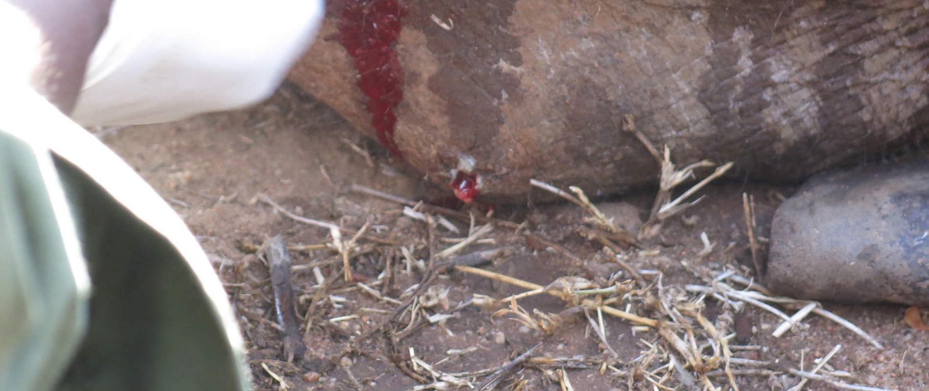
Examination showed a penetrating wound into the soft tissue around the metacarpal bones. This wound was contaminated and oozed pus. On probing with a forceps a bone fracture was confirmed. These findings are consistent with gunshot injuries.


Treatment was achieved by wound debridement with Hydrogen peroxide and cleaning with antiseptic. A long acting antibiotic was injected to control infection spread.
Reversal
Reversal of the anesthetic was achieved by intravenous administration of Diprenophine hydrochloride.
Prognosis
Prognosis for recovery is guarded due to fracture involving the metacarpal bones.
CASE #3: POSTMORTEM OF A BLACK RHINO CARCASS
Date: 5th October 2015
Species: Black Rhino Solo Calf ID. No: 4127
Sex: Male
Age: Juvenile (2.5 years)
Location: Ol Pejeta Conservancy
History
A post mortem examination was conducted on a black rhino carcass at Ol Pejeta Conservancy (OPC) on 5th October 2015. Both the front and rear horns were intact. An examination of the carcass was conducted at the scene of death to document the cause of death and collect genetic samples for RHoDIS.
General observation:
Carcass was in poor body condition (body score 1.5 on a scale of 1-5). Both the front and rear horns had been retrieved for safe keeping and was under custody of the area senior warden. Parts of the carcass had been consumed by scavengers including the perineum, external genitalia, external ear pinna and some viscera.

On flaying the carcass there were subcutaneous bruises and hematomas at the right shoulder area and along the vertebral column. These may have been self inflicted prior to death.
Specific findings
There was extensive autolysis of visceral organs, myocardial hypertrophy, diffuse hemorrhages on the intestinal serosa and fibrinous intestinal adhesions. A purulent mass approximately 15cm in diameter attached to the visceral surface of the spleen was observed and a cut section revealed a ruptured abscess.

Diagnosis
Acute peritonitis secondary to a ruptured splenic abscess.
CASE #4: TREATMENT OF A SICK ELEPHANT
Date: 10th October 2015
Species: Elephant
Sex: Male
Age: Juvenile (5 years)
Location: Naibunga Conservancy, Laikipia
History
This elephant calf in Naibunga Conservancy was reported to have shown severe emaciation over a two week period and had been abandoned by the rest of the herd. It had a swelling on its lower jaw which was oozing pus. Elephant scouts from Space for Giants requested for veterinary evaluation of this calf.
Immobilization, examination and treatment
The elephant was darted from a vehicle after driving it to an open grassland area. Immobilization was achieved using M99 3mg in a single 3cc Dan inject dart and the drugs took effect after 3 minutes.
Examination showed bilateral mandibular abscesses. Irrigation of these abscesses revealed that they penetrated through the root of molar teeth into the oral cavity. Bacterial infection known as Actinomycosis was suspected as the cause.


The abscesses were lanced and lavaged using dilute Hydrogen peroxide and Povidone Iodine. A long acting antibiotic and corticosteroid were also injected.
Reversal
Reversal of the anesthetic was achieved by intravenous administration of Diprenophine hydrochloride.


Prognosis
Prognosis for recovery is guarded due to bone infection which is difficult to manage. Bone tissue is poorly vascularized therefore most antibiotics do not reach bone infection sites.
CASE #5: INJURED ELEPHANT IN DOL DOL
Date: 10th October 2015
Species: Elephant
Sex: Male
Age: Adult
Location: Dol Dol Community Land
History
Livestock herders found this recumbent elephant in a lugga (dry river bed) in Dol Dol. It showed no apparent injury but was believed to have fallen off a cliff at night as it was browsing nearby acacia. There was a steep 12 foot cliff at the bank of the lugga.
Examination and Management
Examination showed paralysis of the hind limbs and loss of sensation of the caudal abdominal muscles and perineum. Traumatic injuries of thoracic spinal cord may have been sustained during the fall.


Efforts to assist it to stand on its feet by roping and pulling with a vehicle were not successful. Due to the extent of its injuries euthanasia was recommended.
CASE #6: WIRE REMOVAL FROM AN ELEPHANT
Date: 11th October 2015
Species: Elephant
Sex: Male
Age: Adult
Location: Lewa Wildlife conservancy (LWC)
History
LWC rangers on patrol reported that a male elephant in a group of 15 elephants was dragging a wire around its left hind limb. This wire which was picked from the fence was coiled around the foot of the elephant. Its herd mates tried to remove the wire by stepping and pulling but instead tightened it.


Immobilization, examination and treatment
This elephant was successfully immobilized using Etorphine hydrochloride. The wire was cut using a wire cutter and removed.
Reversal
Anesthetic drug effect was reversed by intravenous administration of Diprenophine hydrochloride and the elephant fully revived 5 minutes later walked away to join its herd mates.

Prognosis
Good
CASE #7: ARROW HEAD REMOVAL IN ELEPHANT
Date: 12th October 2015
Species: Elephant
Sex: Male
Age: Sub – adult (12 years old)
Location: Samburu National Reserve
History
Scouts from Save the Elephants (STE) reported an elephant which had an arrow head protruding from its back. STE requested for veterinary attention to remove the arrow and treat the wounds.
Immobilization, examination and treatment
Immobilization was achieved using M99 6mg in a single 1.5cc Dan-inject dart from a vehicle with the dart placed at the abdominal muscles. Down time was 8 minutes with the elephant lying on right lateral recumbency.


Examination showed an arrow head penetrating into the dorsal thoracic muscles causing an open abscess.Treatment involved surgical incision to remove the arrow head, lancing the abscess and wound lavage to remove pus. Topical povidone iodine was applied and an antibiotic was injected to control spread of the infection.


Reversal
Complete reversal of anesthetic was achieved three minutes after intravenous administration of Diprenophine.


Prognosis
This young bull is expected to make a complete recovery.
CASE #8: TREATMENT OF A GIRAFFE
Date: 21st October 2015
Species: Reticulated Giraffe
Sex: Male
Age: Adult
Location: El Karama ranch, Laikipia
History
This giraffe had a tight metallic bracelet on its right forelimb causing skin lacerations. As a result the lower limb was swollen. It was first seen on 15th October by wildlife scouts who reported it to ranch management and requested veterinary attention.
Immobilization, examination and treatment
Immobilization was achieved using a combination of Etorphine hydrochloride and Azaperone tartate in a single 3cc Dan-Inject dart from a vehicle. Six minutes later when the giraffe showed signs of sedation it was roped to right lateral recumbency and a blind fold used to cover its eyes.
The steel bracelet was cut using an electric grinder and removed. The metallic object resembled a worn out motor vehicle part which had somehow accidently got stuck on the giraffe’s foot.


Reversal
The anesthetic was reversed by intravenous injection of Diprenophine through the jugular vein. Two minutes later the giraffe was assisted onto its feet by roping around its neck. With the offending object now removed it ran away to join its herd mates.


Prognosis
Good.


