EASTERN CONSERVATION AREA VETERINARY UNIT FEBRUARY 2015 Report by: Bernard Rono SUMMARY This report describes the activities of Meru Veterinary in March 2015
EASTERN CONSERVATION AREA VETERINARY UNIT FEBRUARY 2015
Report by: Bernard Rono
SUMMARY
This report describes the activities of Meru Veterinary in March 2015. Drought experienced in the northern Kenya region has caused serious water and pasture resources scarcity. Many cases reported here is a result of human elephant conflict associated with livestock movement into wildlife conservation areas. We also report ear notching of a white rhino conservancy to enhance rhino monitoring in Lewa and a disease investigation carried out in Grevys’ zebra in Ol Pejeta conservancy.
CASE#1 LAME GIRAFFE
On the 9th of March a giraffe showing lameness and a swollen limb in Meru National Park was reported but a search for the animal was unsuccessful
CASE#2 TREATMENT OF AN ELEPHANT WITH A BULLET WOUND
Date: 12th March 2015
Species: African elephant
Sex: Female
Age: Adult (30yrs)
Location: Suyian Conservancy
History
Wildlife scouts from a lodge in Suyian Conservancy reported that this female elephant had shown severe lameness in the previous 4 days. The right forelimb was swollen with a wound to the lower extremity.


Immobilization, examination and treatment
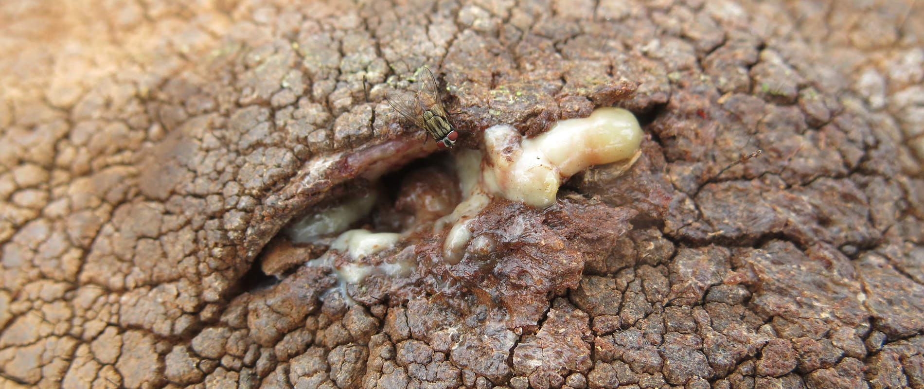
This elephant was located alone near a stream in Suyian and was immobilized using 16mg Etorphine hydrochloride in a 3cc Dan-Inject dart from foot. The elephant went down in 9 minutes. An assessment showed a penetrating wound laterally into the right carpal joint caused by a bullet. The wound was severely infected spreading into the joint causing arthritis.


The wound was lavaged with Hydrogen peroxide and infusion of iodine and antibiotic and anti-inflammatory drug injection given.


Prognosis
Prognosis for recovery is guarded due to spread of infection to the joint.


CASE#3 TREATMENT OF TWO GREVY’S ZEBRA
Date: 13th March 2015
Species: Grevy Zebra
Sex: Female and Male
Age: Adult
Location: Ol Pejeta Conservancy
History
Two Grevys zebra in Ol Pejeta conservancy (OPC) showed progressive loss of body condition and rough hair coat.


The Conservancy recorded mortalities in GZ in January where predominant findings at postmortem included pale mucus membranes, gelatinous fat, complete lack of subcutaneous fat, full stomach and intestines and worms, predominantly Cyathostomes, large Strongyles and tapeworms (Acanthocephala sp).
Animal 1: Immobilization, examination and treatment
The male, approximately 24 months ol,d was anesthetized using a combination of Etorphine and Medetomidine. The drug took a long time to take effect and the zebra had to be re-darted after observing that the first dart may have not discharged.
The male zebra had a heavy tick infestation, a rough hair coat and slightly sunken eyes.
He was given Ivermectin subcutaneous injection and Oxytetracycline intramuscularly. Whole blood and serum blood samples were collected and submitted to the laboratory for analysis.
Reversal
The anesthesia was reversed using a combination of Diprenorphine and Atipamazole. However, the zebra could not stand on his feet. He was given lactated ringer’s solution intravenously but later succumbed at night.
Postmortem Findings
- Slightly pale mucus membrane, tick infestation and the animal had a full stomach.
- There were many small and large strongyles and tapeworms.
- The animal had gelatinous fat and the heart lacked the epicardial fat deposit.
Animal 2: Immobilization, examination and treatment
The adult female was anesthetized using a combination of Etorphine and Xylazine Hcl,
This zebra had a moderate tick infestation. She was in the first trimester of pregnancy with a fair body condition but a rough hair coat. She was given Ivermectin subcutaneous injection.
Complete blood analysis showed abnormal leukocyte distribution with atypical lymphocytosis, anisocytosis, microcytosis and anemia.
Reversal
The anesthesia was reversed using a combination of Diprenorphine and Atipamazole. She could not get up and stand despite several attempted efforts and later succumbed after hitting her head on the ground.
Postmortem Findings
- Tick infestation on the neck, ears and perineum.
- Pale mucus membranes and presence of gelatinous fat in various parts; the animal had petechial streaks on the surface of the heart.
- There was some foam at the bronchial bifurcation with an increased amount of exudation from the lungs.
- The lung tissue was bloody.
- The animal had a 2-3 months old fetus.
- There were strongyles in the stomach lumen.
Conclusion
Both animals had significant tick infestation and a heavy gastrointestinal parasitism. These factors combined with the current lack of quality pasture in the enclosure exacerbate the demand on the animal body.


Capture is a stressor and may have contributed disproportionately to the animals’ lack of energy after reversal of anesthesia and subsequent inability to regain a standing position.
The hematology results in both animals point to normocytic anemia.
It must be noted that anisocytosis may be deficiency induced, as is microcytosis which is normally associated with iron deficiency.
The combination of low albumin and high alkaline phosphatase and aspartate transferase may be due to dehydration or disease and should be investigated further.
CASE#4 RESCUE OF A COLUBUS MONKEY
On the 16th March a Colubus monkey was rescued from a village near Ngaya Forest.


CASE#5 TREATMENT AND EUTHANASIA OF AN ELEPHANT
Date: 16th March 2015
Species: African Elephant
Sex: Male
Age: Juvenile (5 years)
Location: Ol Donyo Nyiro
History
This young elephant was reported to have shown severe lameness affecting the left hind limb. Scouts from the Samburu Trust saw the elephant on 15th March and requested for Veterinary assessment through the KWS outpost in Oldonyo Nyiro. It was in a herd of 26 elephants and its mother had a 1 month old baby.
Immobilization and examination
This elephant was immobilized with 3mg Etorphine hydrochloride in a 1.5cc Dan-Inject dart from a helicopter provided by the Samburu Trust.


An assessment showed swollen thigh muscles of the left hind leg. Manipulation of the femur revealed crepitus and a fracture line. Fractures in elephants have a poor prognosis for recovery, therefore, euthanasia was recommended to relieve pain suffering by this young elephant.


CASE#6 RELOCATION OF A PROBLEMATIC CARNIVORE
On the 16th March a wild carnivore was reported to be causing livestock depradation in community land bordering the park. The carnivore was captured using traps and relocated to the park.

CASE#7 TREATMENT OF AN ELEPHANT WITH A SPEAR WOUND
Date: 17th March 2015
Species: African Elephant
Sex: Female
Age: Adult
Location: Leparua Conservancy
History
Community scouts reported that this elephant had shown little movement in the previous three days. It had a wound which discharged pus on the shoulder and had been left behind by its herd. The scouts requested urgent veterinary treatment.
Immobilization, examination and treatment
This elephant was immobilized using 16mg Etorphine hydrochloride in a 3cc Dan-Inject dart from a vehicle. The dart was placed into the left gluteal muscles and induction time was 5 minutes with the elephant falling onto right lateral recumbency.


Examination showed a spear wound posterior to the right shoulder bone. Probing showed copious amount of pus in the wound.


The wound was thorough washed with Hydrogen peroxide and Povidone iodine applied. Betamox trihydrate 200ml was injected deep intramuscularly and green clay applied to the wound to hasten healing.


Reversal
To reverse the effect of anesthesia 48mg Diprenophine hydrochloride was injected intravenously through superficial veins on the ear pinna. Recovery was smooth and she was reunited with the rest of the herd.


Prognosis
This animal is expected to make a complete recovery in the coming days.
CASE#8 EAR NOTCHING OF WHITE RHINO
Date: 24th March 2015
Species: White Rhino
Sex: Male
Age: 3 years
Location: Lewa Wildlife Conservancy (LWC)
History
Ear notching is one of the key tools in rhino monitoring which aids positive identification of individual rhinos by all observers. On 24th March, Mobile Veterinary units in Meru and Lewa collaborated to ear notch one white rhino in LWC.
Immobilization and ear notching
The white rhino candidate was identified by the rhino monitoring unit in LWC in an open plain which enabled the team to dart the animal from foot. The animal went down in 5 minutes, after which it was blind folded, positioned on sternal recumbency and vital parameters such as rate of respiration recorded.


An appropriate pattern was drawn on each of the two ears and excised using surgical blades. Hemorrhage was controlled using hemostatic forceps and by applying pressure using gauze swabs. Ear notch wounds were liberally sprayed with Oxytetracycline Hcl.


Reversal
Anesthesia was reversed by intravenous administration of 150mg 5% Naltrexone Hcl. Recovery was smooth and this rhino soon joined its companions.


CASE#9 TREATMENT OF AN ELEPHANT WITH A SPEAR WOUND
Date: 25th March 2015
Species: African Elephant
Sex: Female
Age: Adult
Location: Lekuruki conservancy
History
Rangers from NRT in Lekuruki conservancy reported an elephant which showed lethargy, severe emaciation and an infected wound on its abdominal flank. This female elephant which had an 18 month old male calf was in a herd of 20 elephants near Tassia lodge.


Immobilization, examination and treatment
This elephant was darted with 16mg Etorphine hydrochloride 16mg in a 3cc Dan-Inject dart from foot. She was down after 6 minutes after which the calf was chased away.


Examination showed a spear wound on the right flank penetrating into abdominal cavity which on probing with a forceps showed a pus sinus. The wound was thoroughly washed with water, antiseptic and povidone iodine. Betamox trihydrate 200ml was injected intramuscularly and multivitamin injection was also administered.


Reversal
To reverse the effect of anesthesia 48mg Diprenophine hydrochloride was injected intravenously through superficial veins on the ear pinna. Recovery was smooth and she was reunited with the rest of the herd.


Prognosis
This case has a poor prognosis due to its chronic course and severe emaciation.
Acknowledgements
We would like to thank the David Sheldrick Wildlife Trust for providing logistical and financial support to enable treatment of these animals.


