Introduction This report describes activities of the Meru Veterinary Unit in January 2018
Introduction
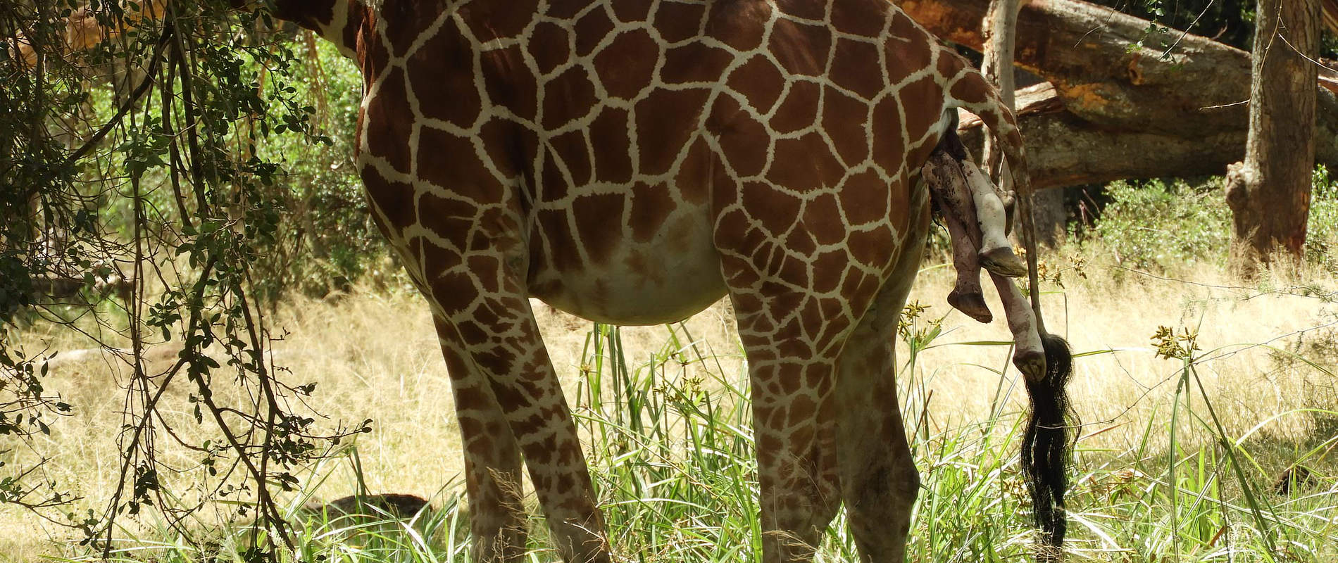
This report describes activities of the Meru Veterinary Unit in January 2018. Among the cases attended were removal of a spear and in an elephant in Namunyak Conservancy and treatment of wounds in an elephant in Milgis. In Meru National Park we treated a common zebra which showed signs of head injury while in Solio ranch we immobilized a reticulated giraffe to relieve obstructed birth.
The unit acknowledges support of the David Sheldrick Wildlife Trust and KWS management for support provided to this unit in providing veterinary intervention to Wildlife in northern Kenya.
CASE 1. TREATMENT OF COMMON ZEBRA
Date: 17 January 2018
Species: Common zebra
Sex: Male
Location: Mulika swamp, Meru National Park
History
We found this zebra during routine patrol in the Park. It showed loss of body condition, circling movements and showed no fear of humans when approached on foot. It had been left behind by its herd mates. We immobilized the zebra to evaluate the cause of illness.
Immobilization and examination
Immobilization was achieved using Etorphine 6mg and Azaperone 80mg in a Dan Inject dart.
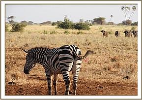
Examination showed bilateral bleeding from the ear canal and circling movements suggestive of traumatic head injury. This can occur due to intra species fight.
Treatment and prognosis
A conservative treatment approach was carried out. Anti – inflammatory drugs and multivitamin injection were administered. This zebra was not found on subsequent search therefore its fate remain unknown.
CASE 2. TREATMENT OF ELEPHANT FOR FIGHT WOUNDS
Date: 23rd January 2018
Species: African elephant
Sex: Male
Location: Pareu, Milgis lugga
History
Scouts from the Milgis Trust reported an elephant had wounds on its left flank as well as above the stifle joint of the right hind limb following a fight with another elephant two weeks earlier.
Immobilization, examination and treatment
The elephant was immobilized using 22mg Etorphine Hydrochloride using a Dan inject intramuscular dart. Darting was done from foot.
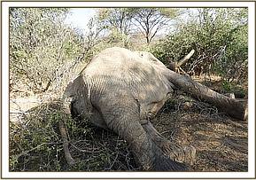

Examination showed deep wounds on the hock joint of the right hind leg. A penetrating wound on the left flank with exuberant tissue was also observed. Wounds are thought to have been as a result of a fight. The wound on the right hind limb was cleaned and exuberant flesh debrided using a scalpel blade, the wound was flushed using dilute hydrogen peroxide and disinfected using tincture iodine soaked swabs. Green clay was smeared on the wound to speed up healing.
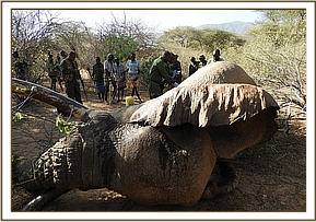

The elephant was treated with 3000mg of Amoxicillin Trihydrate, 100mg of Dexamethasone and 100ml multivitamin intramuscularly. The large male was on left lateral recumbency making it impossible to manage the wounds on the left flank however the parenteral antibiotics administered will help fight infection.
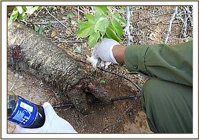

Anesthesia was reversed using 500mg Naltexone Hydrochloride intravenously. The large male’s attempts to get up were futile but it was successfully helped to its feet by help of a vehicle.
Prognosis
Following treatment Milgis Trust scouts reported the elephant had shown improvement and was feeding well. On 30th January, 7 days after treatment, this elephant was found dead on the Milgis lugga. The cause of death may be due to perforating wounds into the left abdominal cavity which was not examined.
CASE 3. TREATING A SPEAR WOUND IN AN ELEPHANT
Date: 24th January 2018
Species: African elephant
Sex: Female
Location: Sarara Lodge, Namunyak Conservancy.
History
The elephant was seen with a spear sticking out of from the right shoulder by Namunyak Conservancy rangers two days earlier.
Immobilization, examination and treatment
The elephant was immobilized using 20mg of Etorphine Hydrochloride and went down on sternal recumbency. Its two herd members were scared off with rifle fire and help of a vehicle before it was positioned on lateral recumbency.


The spear wound penetrated approximately 30cm into the right shoulder muscle. It seems this animal was speared from a cliff. The spear was pulled out and the resultant wound and another wound on the elephant’s right hind limb were flushed with dilute hydrogen peroxide and disinfected using dilute tincture iodine. Green clay was smeared on the wound to speed up wound healing.
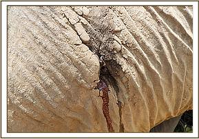

The elephant was treated using 3000mg Amoxicillin Trihydrate and 100mg Dexamethasone administered intramuscularly.


Reversal
Anesthesia was reversed using 60 mg of Diprenorphine hydrochloride intravenously at a prominent ear vein and the elephant was up on its feet 4 minutes later.
Prognosis
Although the wounds were infected, this elephant is expected to make a full recovery following this treatment. Namunyak conservancy rangers will monitor and report if further treatment is required.
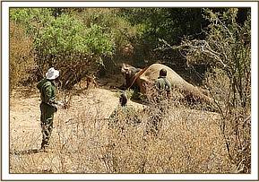

CASE 4. A CASE OF DYSTOCIA IN A RETICULATED GIRAFFE
Date: 31/01/18
Species: Reticulated giraffe
Sex: Female
Age: Adult
Location: Solio Ranch
History
The dam had been spotted by Solio rangers with fetal limbs hanging out from the vulva for 48 hrs.
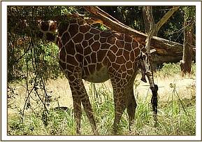

Immobilization and management
The animal was immobilized using 12 mg Etorphine hydrochloride and 80mg Azaperone tartarate combined in one intramuscular dan inject dart and ropes used to cast it down to lateral recumbency. Anesthesia was reversed using 36mg diprenorphine hydrochloride administered intravenously at the jugular.
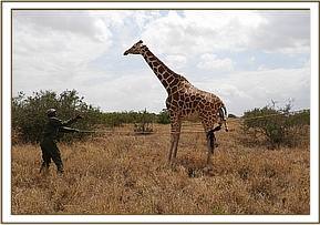

The fetus was in a transverse presentation with all four limbs sticking out of the vulva. The chorioallantois membranes had ruptured depicting that the dam was in stage two of labour.
The transverse presentation was manipulated to a posterior presentation by retropulsion of the forelimbs before applying traction to the hind limbs. The vulva was lubricated with copious amounts of liquid paraffin before attempting to deliver the fetus by traction. The dam unfortunately died during the manipulations and a post mortem was done.
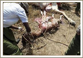

Post mortem findings
The uterine body was lying in the abdominal cavity. In addition to the transverse presentation the fetus also presented with unilateral left hip flexion and ventral flexion of the neck.
The cause of death was prolonged obstructive dystocia due to fetal malpresentation and unilateral left hip flexion causing a lot of fatigue as well as stress from anesthesia.
