EASTERN CONSERVATION AREA VETERINARY UNIT MONTHLY REPORT FEBRUARY 2016 Report by: Bernard Rono INTRODUCTION This report describes the activities of the Meru Veterinary Unit in February 2016
EASTERN CONSERVATION AREA VETERINARY UNIT MONTHLY REPORT FEBRUARY 2016
Report by: Bernard Rono
INTRODUCTION
This report describes the activities of the Meru Veterinary Unit in February 2016. Northern Kenya received little rainfall this month although there is plenty of vegetation and water in many conservation areas following prolonged rains in December/ January.
This unit is supported by the David Sheldrick Wildlife Trust.
CASE #1: ABSCESSES IN ELEPHANTS
Date: 2nd February 2016
Species: African Elephant
Sex: Mixed
Age: Adult
Location: Meru National Park
History
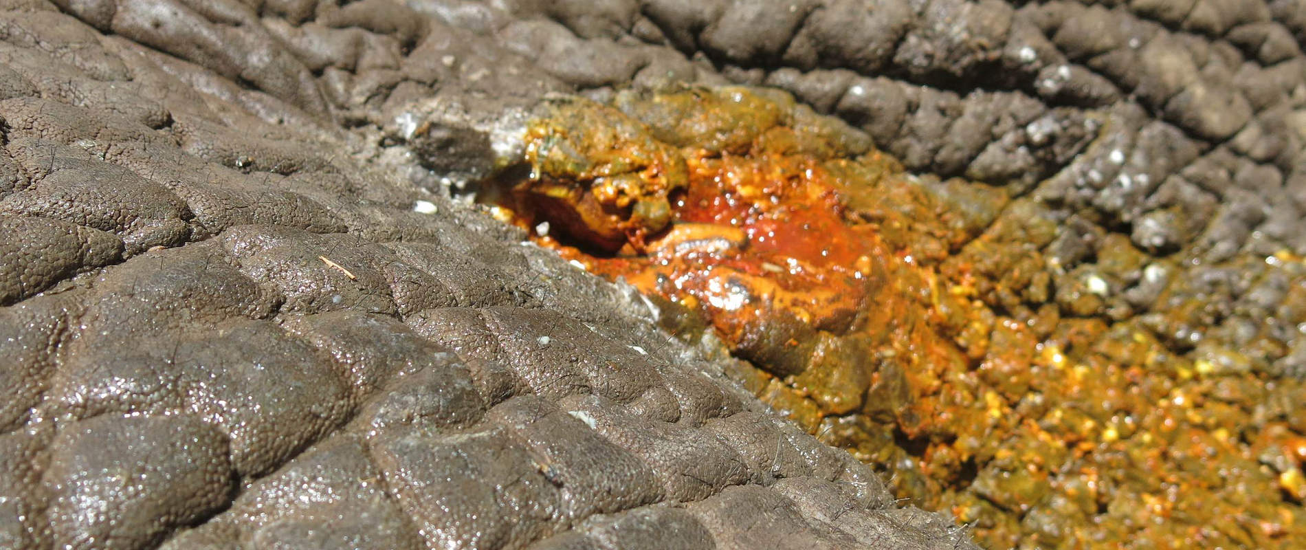
Tour guides from Elsas Kopje lodge in Meru National Park reported that several elephants showed swelling especially in the abdominal area. This followed a period of prolonged rainfall in December/ January hence many elephants had congregated in the park.
Examination
On the 2nd February, we conducted a routine patrol to examine these elephants. Two animals showed cutaneous swellings on the abdominal flank and dorsal to the gluteus. The swellings were benign and diagnosed as abscesses. This condition required no treatment.


CASE #2: CANINE TRYPANOSOMIASIS IN A TRACKER DOG
Date: 3rd February 2016
Species: Canine
Sex: Sub-Adult
Age: Male
Location: Meru National Park
History
On 3rd February, a KWS security dog was reported to have shown reduced appetite and lethargy. Physical examination showed fever and conjunctivae pallor indicative of anemia. A tentative diagnosis of canine ehrlichiosis was made and treatment commenced with oral Doxycyline. After 5 days the dog’s condition deteriorated and it was referred to the small animal clinic at the University of Nairobi for further investigation.
On admission the dog is reported to have shown anorexia, staggering gait and intermittent fever. Later the dog showed muscle tremors, a seizure and finally succumbed. Blood smears showed characteristic trypanosome parasites.
Trypanosomes are transmitted by the tsetse flies (Glossina spp.) which are widely distributed in Kenya. Preferred host for tsetse flies include wild herbivores, although domestic animals including cattle and dogs are occasional hosts. Infected tsetse flies inoculate metacyclic trypanosomes into the skin of animals.
In the dog, control of Trypanosomiasis is primarily through use of prophylactic drugs such as Quinapyramine sulfate. Use of Pyrethrine insecticide wash on the animals is also recommended.
CASE #3: LEOPARD RELEASED IN MERU NATIONAL PARK
Date: 6th February 2016
Species: Leopard
Sex: Adult
Age: Male
Location: Lower Imenti forest
This leopard was reported to have been causing conflict by preying on livestock in lower Imenti forest. A cage trap was set up to capture this animal and it was moved to Meru national park where it was released.


CASE #4: POST MORTEM EXAMINATION OF AN ELEPHANT CARCASS
Date: 17th February 2016
Species: Elephant
Sex: Sub-Adult (15 years)
Age: Male
Location: Lower Imenti forest
History
The warden in charge of Meru station reported that an elephant was recumbent in a farm in lower Imenti. A group of elephants were reported to have raided the maize farm at night and this sub-adult elephant was found recumbent in the morning.
When the Vet Unit visited the farm the team found the elephant had already died and a post mortem examination was carried out to determine the cause of death.
General Examination
Burn marks were seen on the skin which was suspected to have been caused by electrocution. The carcass was significantly bloated and the colon was greatly distended from gas caused by prolonged recumbence. No other significant finding was recorded.
Cause of Death
Cause of death was suspected to be gas colic secondary to an electric shock.
Case #5: Injured elephant in Namunyak conservancy
Date: 24th February 2016
Species: Elephant
Sex: Adult (>40 years)
Age: Male
Location: Namunyak conservancy, Sarara lodge
History
Rangers from the Namunyak Conservancy reported that this elephant had shown severe lameness and a swollen left forelimb. It also had infected wounds which oozed pus.
Immobilization, examination and treatment
Immobilization was achieved using 20mg M99® in a single 3cc Dan-Inject dart from a vehicle. The dart was placed into the gluteal muscles and he went down in 8 minutes. The elephant was then positioned on right lateral recumbency for examination.


Examination showed a stab wound >30 cm deep to the thorax caudal and ventral to the left scapula. These stab wounds were contaminated with maggots and appeared to have been as a result of a fight. The wounds were cleaned with dilute Hydrogen peroxide to remove necrotic tissue and then iodine was applied. Betamox trihydrate, an antibiotic, and Flunixin meglumine, an anti-inflammatory, were administered intramuscularly.


Reversal
For reversal of the anesthetic Diprenophine Hcl was injected intravenously through the superficial ear veins.
Prognosis
Prognosis for this case remains guarded if this animal has internal thoracic injuries.

CASE #6: INJURED COMMON ZEBRA IN LEWA CONSERVANCY
Date: 25th February 2016
Species: Common Zebra
Sex: Adult
Age: Male
Location: Lewa wildlife Conservancy
History
Rangers on patrol in Lewa conservancy reported a common zebra which had expansive wounds to the left gluteus from a lion predation attempt.
Case Management
Observation showed a degloving wound to the left gluteus, severe lameness and listlessness. Due to the extent of these injuries the vet team advised euthanasia.

CASE #7: TREATMENT OF AN INJURED LION IN LEWA CONSERVANCY
Date: 29th February 2016
Species: Lion
Sex: Adult (4 years)
Age: Male
Location: Lewa wildlife Conservancy
History
The Lion Monitoring Team in Lewa Conservancy reported that this lion showed lameness and had wounds on the right hind limb.
Immobilization, examination and treatment
Immobilization was achieved using a combination of Ketamine 300mg and Medetomidine 12mg in a 3cc DanInject dart from a vehicle. The lion went down time in 10 minutes and animal vomited during induction to anesthesia.


Examination showed bruises distal to the hock. Manipulation showed instability of the hock joint which could be as result of injured joint ligaments. This animal was in good body condition and the injuries were most likely caused during a hunt for prey.


The wound was debrided and iodine applied to disinfect the area. Betamox trihydrate 20ml and Dexamethasone injection 20l was injected intramuscularly.
Reversal
The lion was reversed from anesthesia by an intramuscular injection of Antisedan after allowing the ketamine to wear out.
Prognosis
This animal is expected to recover in the next few days.


