EASTERN CONSERVATION AREA VETERINARY UNIT MONTHLY REPORT AUGUST 2016 Summary This report describes activities of the Meru veterinary unit in August 2016
EASTERN CONSERVATION AREA VETERINARY UNIT MONTHLY REPORT AUGUST 2016
Summary
This report describes activities of the Meru veterinary unit in August 2016. During this month the Unit were away on annual leave hence some of the cases were attended by the Sky Vet Unit.
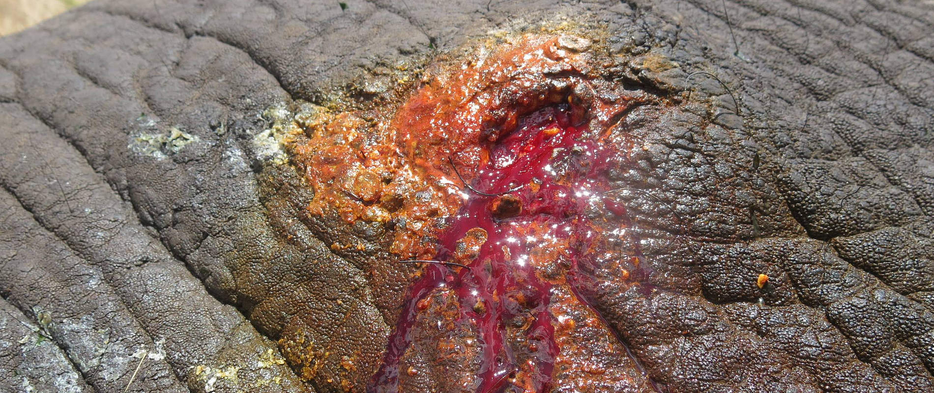
The Meru Vet treated an elephant for an abscess on its flank and offered advice to Ol pejeta Conservancy regarding a black rhino who was reported to have suffered from minor rectal tear.
We would like to thank Dr. Fred Ochieng from KWS headquarters for standing in while we were away. This was made possible through the DSWT Sky Vet program.
CASE#1 SICK BLACK RHINO IN OL PEJETA CONSERVANCY
Date: 27th August 2016
Species: Black rhino
Sex: Male
Age: Adult
Location: Ol Pejeta Conservancy
History and Advice
A black rhino was reported to have passed blood during defecation. This happened on three occasions although it showed normal behavior and good appetite. A minor rectal tear of unknown etiology was suspected. Following discussions with the rhino monitoring team, we recommended further observation of this animal. Two days later it was reported that no more blood was seen in stool.
CASE#2 ABSCESS IN AN ELEPHANT
Date: 30th August 2016
Species: Elephant
Sex: Male
Age: Adult
Location: Meru national park
History
This elephant was found during a patrol in the park. It had pus oozing from a swelling in the abdominal flank. This elephant was darted for examination and treatment.


Immobilization, examination and treatment
Immobilization was achieved using Etorphine hydrochloride 18mg in a single 3cc Dan-Inject dart with a 2.2 × 60 mm needle. Darting was done from a vehicle with the dart placed into the gluteal muscles. After 10 minutes this elephant went into left lateral recumbence.


Examination showed an open abscess. Debridement of the abscess was carried out to remove pus and necrotic tissue using hydrogen peroxide and iodine. Green clay was then applied and a systemic antibiotic was injected to prevent spread of infection.


Prognosis
This animal will make a full recovery in the coming days.


CASE#3 REMOVAL OF A LION COLLAR
Date: 31/08/2016
Species: Lion
Sex: Female
Age: Adult
Location: Lewa conservancy
History
A female lion in Lewa conservancy was immobilized to remove a tracking collar which had malfunctioned. The lioness was also treated for lameness on its right forelimb.
Immobilization, collar removal and treatment
The lioness was darted using a combination of Ketamine hydrochloride and Medetomidine Hcl from a vehicle with the dart placed into the shoulder muscles. Down time was 6 minutes.
A blindfold was applied and the lion moved to a shade for examination and collar removal.

The lion also had a wound on the medial claw of the right forelimb. The hair was clipped from around the wound so it could be cleaned. Povidone iodine and topical antibiotic spray were also applied.
Reversal
Antisedan was injected intramuscularly 45 minutes after sedation to reverse the effects of medetomidine Hcl. Ten minutes later the lioness came around.


