EASTERN CONSERVATION AREA VETERINARY UNIT MONTHLY REPORT APRIL 2018 Report by: Bernard Rono, Veterinary Officer SUMMARY This report describes the activities of the Meru veterinary unit operating in northern Kenya in April 2018
EASTERN CONSERVATION AREA VETERINARY UNIT MONTHLY REPORT APRIL 2018
Report by: Bernard Rono, Veterinary Officer
SUMMARY
This report describes the activities of the Meru veterinary unit operating in northern Kenya in April 2018. Northern Kenya experienced above average rainfall during this month with a rejuvenation of vegetation and water sources in the wildlife park. However, most parts of the park were inaccessible for wildlife monitoring and tourism with few cases of injuries reported. One major activity carried out in Meru National Park Sanctuary was rhino ear notching. This was aimed at individual identification of rhinos. The exercise was also timed to coincide with the extension of the rhino sanctuary to provide a wider area for establishment of rhino territories. A black rhino was also treated for cutaneous wounds caused by filariae in Meru park while a common zebra which showed lameness was immobilized to investigate the cause of lameness. These cases are described in detail below.
We would like to acknowledge support of the DSWT which provides financial and logistical support to the Meru veterinary unit and the KWS management for facilitating the work of this unit in northern Kenya.
CASE #1: RHINO EAR NOTCHING IN MERU NATIONAL PARK
Species: Black rhino and White rhino
Location: Rhino Sanctuary in Meru National Park
Date: April 3 - 10, 2018
Background information
Two (2) black rhinos and fifteen (15) white rhinos were captured and ear notched in Meru national park 3 - 10 April, 2018. This exercise aimed to identify individual rhinoceros by specific ear-notch pattern and a particular name and insert microchips in both horns, neck and rear region. Ear notching is one of the key tools in rhino monitoring which aids positive identification of individual rhinos by all observers. The current exercise was carried out by KWS personnel from the Rhino Program office, Veterinary and Capture department, Air wing, Research department in Meru and DSWT/KWS Meru mobile veterinary unit.
Ear notching methodology
i.Rhinoceros candidate selection and capture of rhinoceros
Rhinos selected for ear notching were individuals two to four years old that had not been notched previously (described as clean animals). This selection criterion was used to minimize risk associated with post immobilization separation of calves which are dependent on their mother for nutrition and nurture.
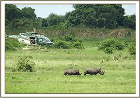
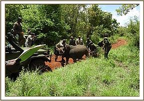
The process started by conducting an aerial search for the suitable candidates by experienced spotters using both a fixed wing plane and a helicopter. Candidates were identified on the basis of their territories, individual attributes such as age, size and sex and group attributes for instance number in a group or identity of its companions. After the target being positively identified, it was first driven to open ground and darted from a helicopter.
ii.Anesthesia reversal
All the rhinos were darted from a helicopter using a Dan-inject rifle, two (2) milliliter darts with a 2.2 × 60mm needle. Etorphine Hcl (Captivon®0.98%) and Azaperone tartate combination was used for anesthesia.

The anesthesia was reversed in white rhinos by intravenous injection of 5% Naltrexone Hcl followed by Diprenorphine Hcl (Activon® 1.2%) given by intramuscular route. Anesthesia in black rhinoceros was reversed using Diprenorphine Hcl given by intravenous route and a ¼ of the dose by intramuscular route.
iii.Physical examination and monitoring of anesthetized rhinos
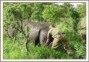
After being immobilized, the ground team moved in quickly to process the animal. Immobilized rhinos were positioned on sternal recumbency with neck extended to ensure patent airway. Vital parameters were monitored including respiration rate and depth, colour of mucous membrane, and rectal body temperature. In addition rhinos showing signs of respiratory distress were treated with Butorphanol tartrate (1%) 0.5 ml and Doxapram 20mg administered intravenously. To control hyperthermia immobilized animals were doused with cold water. Animals were blind-folded to reduce capture stress. All immobilized rhinos were examined physically for injuries, wounds and filarial worms wound lesions and treated appropriately. For control of ectoparasites all immobilized rhinos were treated with an ectoparasiticide pour on.

iv.Ear-notching technique, fitting transponders and Sample Collection
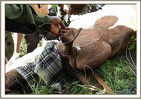
A predetermined pattern was excised on the ears of each of the anesthetized rhinos using surgical scissors and blades. Hemorrhage was controlled using hemostatic forceps and by applying pressure using gauze swabs. Ear notch wounds were liberally sprayed with Oxytetracycline Hcl.
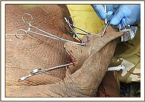

Four transponders (microchips) were fitted to the animal; 1 in each horn, 1 in the neck region and another at the base of the tail. However, the microchips could not be fitted in the rear horn of most of the white rhinos as the horns were too small to be drilled. After fitting, the microchips were checked to be working using a scanner and the respective codes recorded in a data sheet.

Conclusion
Ear notching is a reliable and easy method for identification of rhinos in the wild. In this case all target white rhino candidates were captured and ear notched. Two black rhino out of seven target animals were ear notched during this period. Due to heavy rains experienced in April and thick vegetation, parts of the sanctuary which are predominantly black rhino territories were inaccessible. Hence we recommend a follow up exercise in September during the dry season to target black rhino.
CASE #2: FILARIOSIS IN A BLACK RHINO
Date: 8th April 2018
Species: Black Rhino
Sex: Male
Location: Meru National Park
History
The black rhino had a large superficial wound on the left hind quarter and deep cutaneous wounds on the dorsal thoracic area. The animal was spotted during the ear notching exercise and was immediately immobilized for treatment to prevent further enlargement of the wound.
Immobilization, examination and treatment
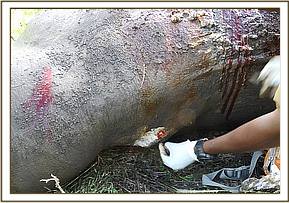
The animal was immobilized using Etorphine hcl 4mg and Azaperone tartarate 50mg combined in one 1.5ml Dan-inject intramuscular dart from a helicopter. The rhino had deep cutaneous wounds on its right flank and left flank and on the left rump approximately 15cm in diameter which was healing. The wounds were characterized by erosive ulceration and crust formation.

Samples of affected skin were collected for histopathological examination before the wounds were cleaned and debrided using dilute hydrogen peroxide then rinsed with dilute tincture iodine. Green clay was smeared on the wounds to hasten healing and then they were covered adequately with Oxytetracycline aerosol. The animal was treated with Ivermectin 200mg subcutaneously and Amoxicillin trihydrate 12000mg intramuscularly.

Reversal and Prognosis
The animal was revived using 50mg Naltrexone hcl intravenously at the ear vein and 12mg diprenorphine intramuscularly. Wounds caused by filariae respond well to ivermectin treatment. We advised the rhino monitoring team to follow up and report on the progress of this rhino.
CASE #3: LAMENESS IN A COMMON ZEBRA
Date: 18th April 2018
Species: Common zebra
Sex: Female
Location: Meru National Park
History
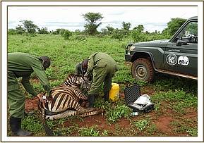
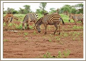
This adult female zebra was lame on its left hind limb with slight swelling around the pastern joint. The animal showed signs of pain and discomfort. On trot this zebra showed weight shifting lameness favoring the affected limb.
Immobilization, examination and treatment
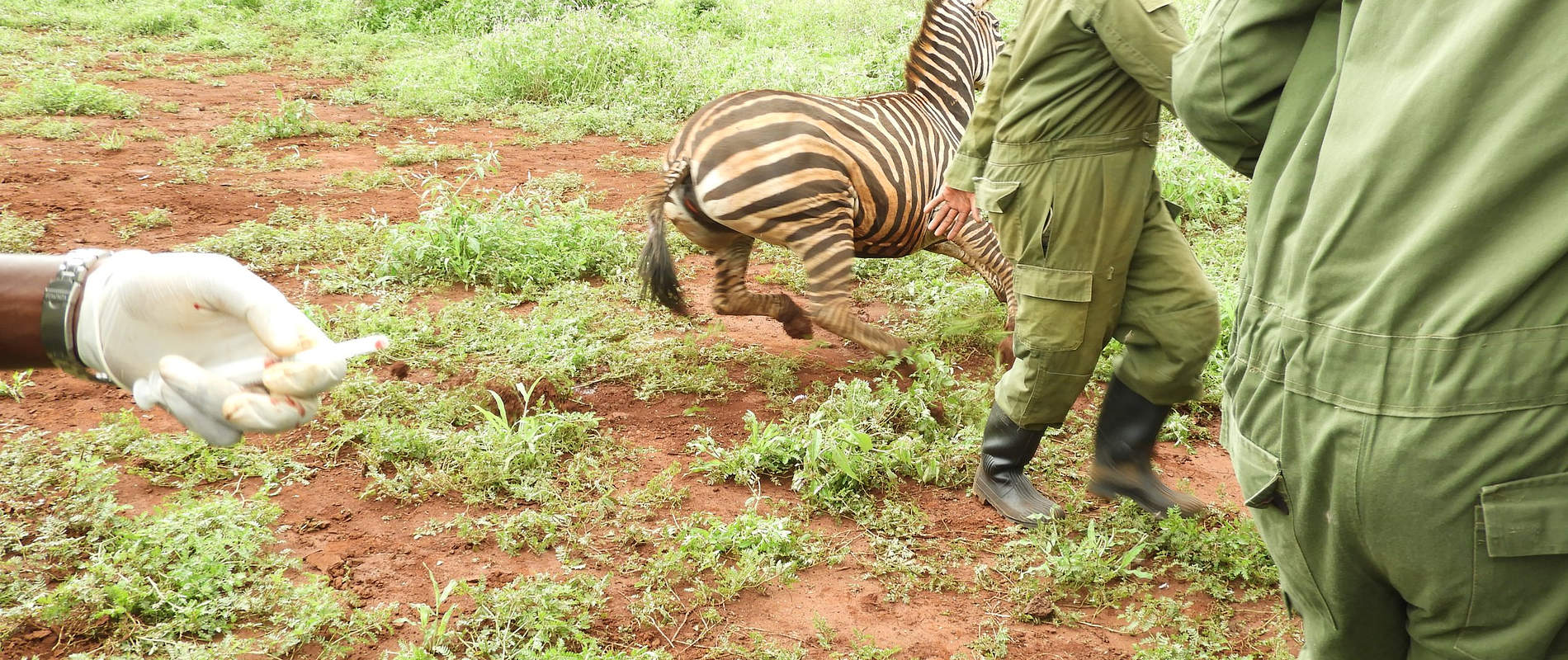
The zebra was immobilized using Etorphine Hydrochloride 5mg and Azaperone tartarate 80mg combined in a 1.5ml Dan-inject intramuscular dart and went down on lateral recumbency. The affected limb was slightly swollen with no snare injury. Joint manipulations and deep palpations showed a sprain. The zebra was treated with Amoxicillin trihydrate 6000 mg and flunixin meglumine 500mg administered intramuscularly.

Reversal
The anesthesia was reversed using 18mg of Diprenorphine hydrochloride intravenously at the jugular vein and the animal was up on its feet a minute later. As this was a mild injury to the leg, the prognosis for complete recovery is good. We expect that the zebra will soon regain good health.
