EASTERN CONSERVATION AREA VETERINARY UNIT MONTHLY REPORT Report by: Bernard Rono, Veterinary Officer Summary This report describes the activities of the Meru veterinary unit in northern Kenya in April 2016
EASTERN CONSERVATION AREA VETERINARY UNIT MONTHLY REPORT
Report by: Bernard Rono, Veterinary Officer
Summary
This report describes the activities of the Meru veterinary unit in northern Kenya in April 2016. Many parts of northern Kenya received rainfall in April, therefore adequate browse and water was available for wildlife.
In Ol Jogi wildlife conservancy a female giraffe with an obstructed birth was treated. In El Karama ranch an elephant showing severe lameness was attended to while in Meru national park we treated a white rhino for cutaneous filariasis.
This unit is supported by the David Sheldrick Wildlife Trust.
CASE #1: DYSTOCIA IN A RETICULATED GIRAFFE
Date: 10th April 2016
Species: Reticulated giraffe
Sex: Female
Age: Adult
Location: Ol Jogi ranch
History
A female giraffe in Ol Jogi Wildlife Conservancy was seen with a dead fetus in its birth canal for two days. Veterinary intervention was required to save the giraffe from severe endotoxemia as it was unable to push the fetus out.
This giraffe was raised at an orphanage in the conservancy and later released into the wild. It was a first time mother.
Immobilization, examination and treatment
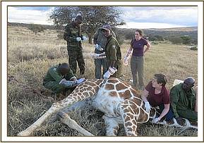
Immobilization was achieved using a combination of Etorphine hydrochloride 10mg and Azaperone tartate 40mg in a 1.5cc Dan-Inject dart from foot. Five minutes later the giraffe was roped to lateral recumbency.
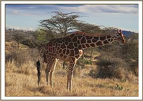

Genital examination showed a dead fetus on anterior longitudinal presentation and dorsal - sacral position. The extended head, neck and front limbs were protruding from the vulva. Fetal – maternal disproportion (small birth canal) was suspected as the cause of dystocia.


Liquid paraffin was applied liberally to lubricate the birth canal. Ropes were applied in loops on the distal metacarpus to aid in traction. The fetus was manipulated gently and manual traction applied caudally and ventrally. A fully formed dead male fetus was delivered.
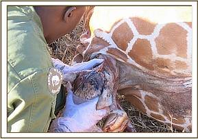

Fetal membranes were trimmed from the uterus and Oxytetracycline pessaries administered. Parenteral antibiotic and analgesic drugs were also injected.
Prognosis
This giraffe was reported to have recovered one week after treatment. It is expected that this giraffe will be able to breed in future.

CASE #2: CUTANEOUS FILARIASIS IN A WHITE RHINO
Date: 12th April 2016
Species: White rhino
Sex: Male
Age: Adult
Location: Meru national park
History
A male rhino in Meru National Park was reported to have shown a large cutaneous wound on the abdomen which required treatment. The wound had grown progressively over a period of two weeks.
Immobilization and examination
The rhino was immobilization with a combination of 5mg Etorphine and 60mg Azaperone in 1.5cc Dan-Inject dart from a helicopter. The rhino was darted in the left rump and induction time was 7 minutes.
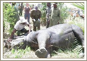

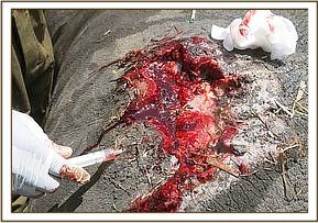
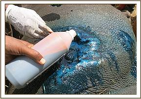
Examination showed ulcerative cutaneous wounds with serrated edges and pockets of pus. Wounds emitted a foul smell due to tissue necrosis caused by bacterial infection. These wounds are characteristic of cutaneous filariasis. The wounds were thoroughly washed with water diluted with hydrogen peroxide and then the necrotic tissue was cut away. Tincture of Iodine soaked in gauze swabs was applied and 1% Ivermectin 300mg and 20% Oxytetracycline administered subcutaneously and intramuscularly.

Reversal
Anesthesia effects were reversed within 3 minutes of intravenous injection of Naltrexone Hcl 150mg.
Prognosis
Filarial wounds respond to Ivermectin and antibiotic treatment, therefore this rhino is expected to make a speedy recovery. The rhino monitoring team will observe and report on their progress.

CASE #3: POST MORTEM EXAMINATION OF A RHINO
Date: 16th April 2016
Species: Black rhino
Sex: Female
Age: Adult
Location: Ol Pejeta Conservancy
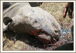
A female black rhino carcass was found in Ol Pejeta Conservancy on 16th April 2016. Both horns had been removed by suspected poachers. A post mortem examination of the carcass showed that it had several gunshot wounds to its chest. Severe internal hemorrhage was also observed in the thoracic and abdominal cavities. Projectiles consistent with small caliber fire arms were removed from the colon, pectoral muscles and the muscles of the forelimb. A female fetus in first trimester gestation was found in the uterus. Our findings showed that this black rhino died from severe hemorrhage due to gunshot injuries.

CASE #4: LAMENESS IN ELEPHANT
Date: 25th April 2016
Species: Elephant
Sex: Male
Age: Adult
Location: El Karama ranch
History
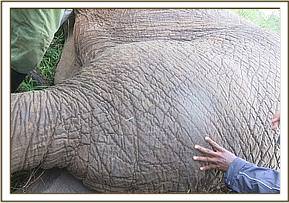
This elephant showed severe lameness and a swollen left thigh. It was first seen on 24th April and treatment was carried out one day later.
Immobilization was achieved using 18mg Etorphine in a 3cc Dan-Inject dart from foot. Induction time was eight minutes after which the elephant fell onto sternal recumbency. The elephant was roped to right lateral recumbency for examination.


Examination and management
The left thigh was grossly enlarged and warm to touch. Crepitus was felt on manipulation of the limb. A tentative diagnosis of fracture of the femur was made. An attempt to radiograph this leg was not successful due to huge muscle mass. Conservative treatment was attempted using parenteral antibiotics and anti – inflammatory drugs.

Reversal
To reverse the effect of anesthetic drugs Diprenophine Hydrochloride was injected intravenously. After three minutes the elephant was assisted to standing position. However excessive mobility and leg deformity was later noted when this elephant walked off which confirmed a fracture and euthanasia was arrived.
Post mortem findings
There was a hematoma affecting the muscles of the femur and comminuted fracture of the femur with severe bone displacement.
