FIELD VETERINARY REPORT FROM MASAI MARA – JUNE 2018 By Campaign Limo Introduction: This past month we have seen a slight drop in precipitation with plenty of food and water still available to the wildlife
FIELD VETERINARY REPORT FROM MASAI MARA – JUNE 2018
By Campaign Limo
Introduction:
This past month we have seen a slight drop in precipitation with plenty of food and water still available to the wildlife. There was a drop in cases reported and handled during the month with one unfortunate incident which lead to the loss of a big elephant within the ecosystem.
The following cases were handled during the month:
CASE #1 DE-SNARING OF AN ELAND
Date: 7th June 2018
Species: Eland
Sex: Female
Age: Adult
Location: Olerai Conservancy, Masai Mara
History:
This adult female eland was seen dragging a long plain wire firmly attached to her left hind leg by the Olerai Conservancy team who immediately notified the Mobile Veterinary unit for intervention. She was found with a mixed herd of about twenty elands with the plain wire firmly attached to her left hind leg. Upon approach, she seemed to be a bit shy but in good health despite her injuries.
Immobilization, examination and treatment:
Restraint was achieved chemically by use of a combination of 10mgs Etorphine hydrochloride and 70mgs Azaperone tartarate delivered through a 1.5ml Daninject dart. Darting was carried out from a vehicle. She took off after being darted with the loose and longer portion of the snare snapping off as she fled. She remained with the tighter portion round her leg. The drugs took full effect after ten minutes with the eland assuming left lateral recumbency.


Examination revealed a tight plain wire snare on her left hind leg slightly above the hock joint which had created some soft tissue injuries but was not infected.The wound was cleaned with copious amounts of water, debrided with the help of hydrogen peroxide, rinsed with clean water and disinfected with tincture of iodine.Topical oxytetracycline wound spray was then applied. Other treatments given include parenteral administration of Amoxicillin antibiotics and Flunixin meglumine anti-inflammatories.


Reversal and Prognosis:
The anaesthetic was reversed by intravenous administration of 30mgs Diprenorphine hydrochloride delivered through the jugular vein. She woke up after three minutes to join her fellow grazers. Her prognosis for recovery is good.

CASE #2 POST MORTEM OF A DEAD VULTURE
Date: 14th June 2018
The carcass of this vulture was presented for examination to the KWS Research Station by members of the Peregrine Fund. It was retrieved from a tree close to Olarro Conservancy.
General observation;
- This was a two to three day old carcass beginning to putrefy.
- She was found to be a female and appeared to have been in fair body condition before death.
- The crop had already been opened and was almost empty.
- She was considered a mature adult.
On opening the carcass, the following was noted;
- She had fair muscle and fat cover.
- The liver, being a metabolically active organ, was undergoing autolysis. This was considered a normal occurrence considering the time after death.
- No internal injuries were detected apart from the already opened crop.
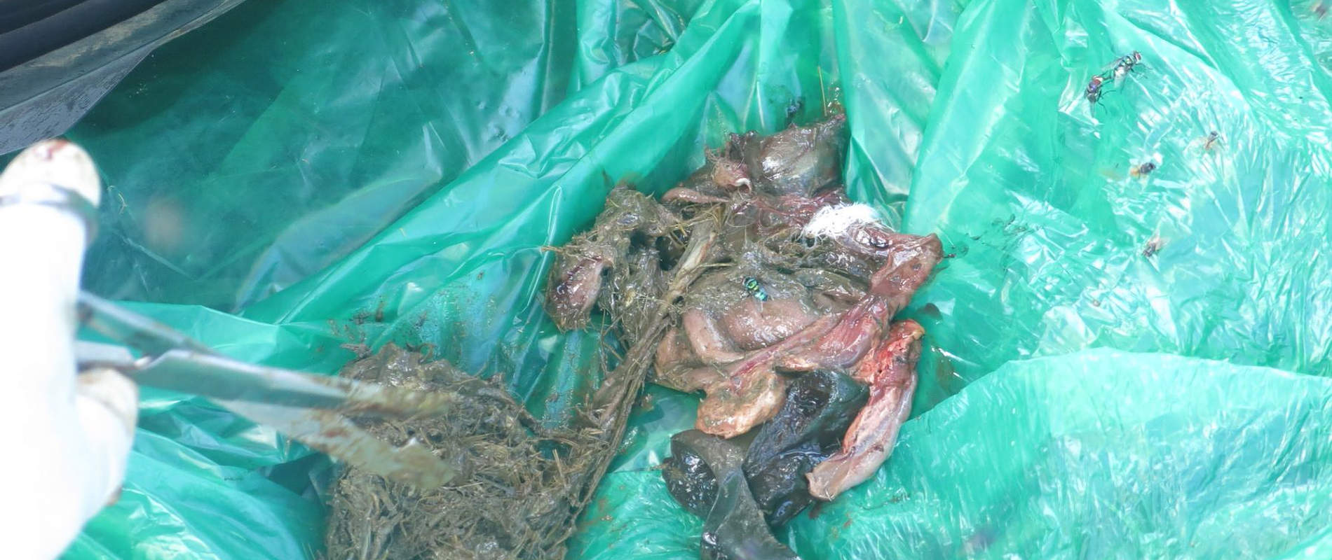
- Of interest, were the stomach contents which comprised almost exclusively of long dry grass forming a big ball.
- The lower gastrointestinal tract beyond the stomach was devoid of any solid matter. Much of the contents were fluidy though scanty. No fully formed faecal matter was traced.
Conclusion:
This vulture could have died due to impaction by phytobezoars. The finding of the excessive and almost exclusive grass ball within her stomach was considered abnormal given that vultures are flesh eaters. The ball had occupied much of the space in the stomach occluding the exit to the lower gastrointestinal tract with attendant associated complications, such as starvation due to gastrointestinal function interference.


CASE #3 EXAMINATION OF A DEAD ELEPHANT
Date: 20th June 2018
Species: African elephant (Loxodanta africana)
Sex: Male
Age: 40 – 45 years
Location: Kichwa Tembo area
History:
This carcass was seen the morning of the 20th of June, still intact, close to a valley near Kichwa Tembo, and was reported to the Veterinary unit by Mara Triangle Conservancy management. The unit was requested to complete an autopsy in order to determine the cause of death of this hither to healthy elephant. It was reported that this elephant was known to be friendly to humans and was commonly sighted in this area.
General Observation:
He was found dead and lying on his right lateral side with the following general findings;
- He appeared to have been in perfect body condition with a body score of 4.5 out of 5 (5 being perfect and 1 being poor).
- Both tusks were intact.
- The carcass was still fresh (no rigor mortis), estimated to be less than 12 hours old.
- There was an old scar on his left rump.
- Scavengers had pulled out part of flesh around the left jaw and the tip of the trunk. Anal tissue and the right teat had also been scavenged on.
- Clotted blood was seen within the mouth and some was attached to the hard palate.
- A penetrating wound was seen behind the left ear through the ear pinna, with the pinna cartilage being shattered. This was accompanied with a lot of bleeding.
- It appeared like there was little struggle at the scene before death with tree shrubs around the area trampled on as the elephant was going down.
- Blood stains were seen at the scene of death, limited to a radius of four meters from the carcass.
- When the carcass was turned over, no cutaneous discontinuity was observed on the right side.


Specific findings:
- There was excessive bleeding with suggestive deep seated injury and massive tissue damage. The source of the bleeding was the penetrating wound just behind the left ear. This opening had a diameter of about 2 inches.
- The course of the wound accessed from the left side was ventro-medial with several bone fragments were retrieved. This opened up into the left upper jaw with the first molar being shattered, together with the left jawbone and left side of the hard palate. Among the bone tissues retrieved were skull bone fragments and displaced hyoid bone. This led to the bleeding seen from the mouth.
- The opening between the entry wound and exit wound, and the skull bone tissue recovered, meant the left side of the brain was damaged and this effectively stunned this elephant.


Conclusion:
This elephant died from injuries caused by a high calibre projectile through the left side of his brain. Entering through the left jaw, and exiting dorso-laterally just behind the left ear. As the projectile exited, the left ear pinna was also damaged and its cartilage fragmented. With severe bleeding and brain damage, this elephant died with little struggle within a short radius of being injured. The circumstances point to human involvement which resulted in the death of this big elephant.
CASE #4 INJURED ELEPHANT
Date: 26th June 2018
Species: African elephant
Sex: Male
Age: Adult
Location: Olarro Conservancy
History:
Whilst on their normal patrols the Olarro rangers spotted this bull, in the company of two other young bulls, with a suppurating wound on his right flank. They immediately called the mobile Veterinary Unit for intervention.
General observation:
The three bulls were found calmly browsing near a small thicket. A small discharging wound could be seen on the right flank of this big bull.
Immobilization, examination and treatment;
Restraint was achieved chemically through darting the bull with 15mgs Etorphine hydrochloride delivered through a 1.5ml Daninject dart. Darting was done from a vehicle. It took ten minutes for the drugs to take full effect with this elephant assuming right lateral recumbency in an open area. He was rolled over to expose his injury. This was a three to four day injury likely caused by a non-poisoned arrow which had since fallen, as probing and scanning with a metal detector revealed that there was no foreign body in the wound.


All necrotic tissues were removed with the help of hydrogen peroxide for debriding. A copious amount of water was used to rinse the wound before tincture of iodine was applied as disinfectant. Topical Oxytetracycline spray was lastly applied. Other treatments carried out included parenteral administration of Amoxicillin antibiotic and Flunixin meglumine anti-inflammatory.


Reversal and Prognosis:
Reversal was achieved through the administration of 36mgs Diprenorphine Hydrochloride via a prominent ear vein. He woke up in four minutes, stood calmly for a while before joining his colleagues. His prognosis for recovery is good.


Conclusion:
The Mara Mobile Veterinary Unit would like to extend its gratitude to all the stakeholders who helped in reporting and locating wild animals that required medical attention during this month. Thanks to the Minara foundation through DSWT and KWS management for their continued partnership which has helped the unit reach and treat many wild animals under distress.


